
Ultrasound Pelvis TVs
Transvaginal ultrasound (TVS) of the pelvis is a specialized imaging procedure used to visualize the pelvic organs, including the uterus, ovaries, fallopian tubes, and surrounding structures, in females. Unlike abdominal ultrasound, which is performed externally over the abdomen, transvaginal ultrasound involves inserting a small ultrasound probe into the vagina for closer and clearer imaging of the pelvic organs. This procedure is commonly used in gynecology for various diagnostic purposes, including assessing pelvic pain, evaluating fertility issues, monitoring pregnancies, and detecting gynecological conditions.
Purpose of Transvaginal Ultrasound (TVS) of the Pelvis
Evaluation of Uterine Structures
Transvaginal ultrasound allows for detailed visualization of the uterus, including its size, shape, position, and internal structures such as the endometrium (inner lining) and myometrium (muscular wall). It helps detect abnormalities such as fibroids, polyps, adenomyosis, and congenital uterine anomalies.
Assessment of Ovaries and Follicles
TVS provides high-resolution imaging of the ovaries, enabling the detection of ovarian cysts, tumors, and other abnormalities. It also allows for monitoring of ovarian follicle development during the menstrual cycle and assessment of ovarian reserve in fertility evaluations.
Diagnosis of Gynecological Conditions
Transvaginal ultrasound is instrumental in diagnosing various gynecological conditions, including ovarian cysts, endometriosis, pelvic inflammatory disease (PID), ovarian torsion, and ectopic pregnancy. It helps identify the location, size, and characteristics of pelvic masses and assists in treatment planning.
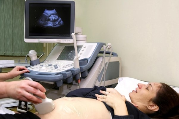
Procedure of Transvaginal Ultrasound (TVS)
Preparation and Process
The transvaginal ultrasound procedure typically involves the following steps:
Patient Preparation: The patient is positioned on an examination table in the lithotomy position, similar to a pelvic exam. A specialized transvaginal ultrasound probe is covered with a disposable sheath and lubricated before insertion.
Probe Insertion: The ultrasound probe is gently inserted into the vagina and maneuvered to obtain optimal views of the pelvic organs. The patient may experience mild discomfort but should not feel significant pain during the procedure.
Ultrasound Examination: The ultrasound technician or healthcare provider obtains real-time images of the pelvic organs by moving the probe to visualize different angles and structures. Measurements of organ size, dimensions, and any abnormalities are recorded.
Image Acquisition: The ultrasound machine displays the images on a monitor for interpretation. The technician may take screenshots or video clips of specific findings for documentation and analysis.
Clinical Applications
Fertility Assessment
Transvaginal ultrasound is used in fertility evaluations to assess ovarian function, follicle development, and the thickness of the endometrium. It helps identify potential causes of infertility, such as ovarian disorders, uterine abnormalities, or tubal blockages, guiding treatment decisions for assisted reproductive technologies (ART).
Pregnancy Monitoring
During early pregnancy, transvaginal ultrasound is used to confirm pregnancy, visualize the gestational sac, and assess fetal viability. It helps determine gestational age, detect multiple pregnancies, and diagnose conditions such as miscarriage or ectopic pregnancy. TVS is also used for monitoring fetal growth and development during the first trimester.
Advantages and Limitations
Advantages: Transvaginal ultrasound provides high-resolution imaging of the pelvic organs with improved clarity and detail compared to abdominal ultrasound. It offers better visualization of structures close to the vagina, such as the ovaries and uterus, and is less affected by bowel gas interference. TVS is well-tolerated by most patients and does not require a full bladder for imaging.
Limitations: While transvaginal ultrasound is highly effective in visualizing the pelvic organs and detecting many gynecological conditions, it may have limitations in detecting abnormalities located outside the vagina or uterus. It may also be less comfortable for some patients compared to abdominal ultrasound, particularly those with vaginal or pelvic pain.
Integration with Healthcare
Transvaginal ultrasound is routinely integrated into gynecological practice for the evaluation and management of various pelvic conditions and fertility issues. It complements other diagnostic tests, such as pelvic exams, blood tests, and hysterosalpingography, to provide a comprehensive assessment of gynecological health. Results of TVS guide treatment decisions, monitoring strategies, and follow-up care tailored to the individual patient’s needs. Regular imaging may be recommended for ongoing surveillance and disease management.