
Ultrasound of Abdomen and Pelvis
Abdominal and pelvic ultrasound is a diagnostic imaging procedure used to visualize the structures within the abdomen and pelvis using high-frequency sound waves. It provides detailed images of organs such as the liver, gallbladder, pancreas, kidneys, bladder, uterus, and ovaries, helping diagnose various medical conditions and guide treatment decisions. Abdominal and pelvic ultrasound is non-invasive, safe, and widely used in clinical practice.
Purpose of Abdominal and Pelvic Ultrasound
Evaluation of Abdominal Organs
The primary purpose of abdominal ultrasound is to assess the anatomy and function of abdominal organs, including the liver, gallbladder, pancreas, spleen, kidneys, and abdominal aorta. It helps detect abnormalities such as liver masses, gallstones, pancreatic cysts, renal cysts, and abdominal aortic aneurysms.
Assessment of Pelvic Organs
Pelvic ultrasound is used to visualize pelvic organs in both males and females, including the bladder, uterus, ovaries, and prostate gland. It helps diagnose conditions such as ovarian cysts, uterine fibroids, endometrial thickening, pelvic inflammatory disease (PID), and benign prostatic hyperplasia (BPH).
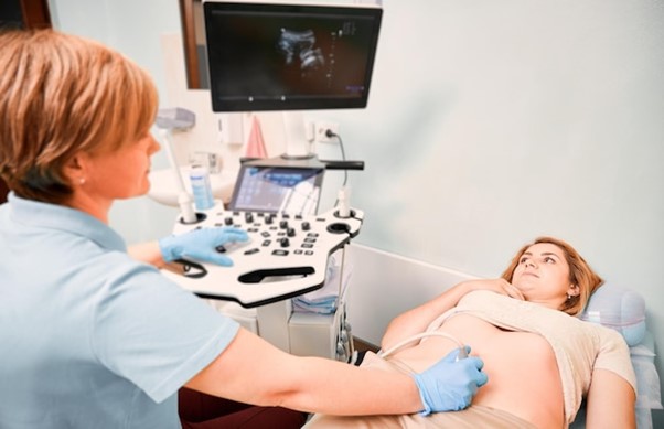
Procedure of Abdominal and Pelvic Ultrasound
Preparation and Process
The abdominal and pelvic ultrasound procedure typically involves the following steps:
Patient Preparation: The patient may be asked to fast for a few hours before the test, particularly if the liver or gallbladder is being evaluated. A full bladder may be required for pelvic ultrasound in some cases.
Positioning: The patient lies on an examination table, and a water-based gel is applied to the skin overlying the abdomen and pelvis to facilitate ultrasound transmission.
Ultrasound Examination: A handheld ultrasound transducer is moved over the skin in the abdominal and pelvic regions to obtain images of the organs. The technician may use different scanning techniques to visualize each organ thoroughly.
Image Acquisition: The ultrasound machine captures real-time images of the abdominal and pelvic organs, displaying them on a monitor for interpretation. Measurements of organ size, dimensions, and any abnormalities are recorded.
Clinical Applications
Diagnosis of Abdominal Conditions
Abdominal ultrasound is used to diagnose a wide range of abdominal conditions, including liver cirrhosis, hepatitis, cholecystitis, pancreatitis, renal cysts, hydronephrosis, and abdominal masses. It provides valuable information about organ size, shape, texture, and blood flow, aiding in the diagnosis and management of various medical conditions.
Evaluation of Pelvic Disorders
Pelvic ultrasound is instrumental in diagnosing gynecological and urological conditions affecting the pelvic organs. It helps identify abnormalities such as ovarian cysts, uterine fibroids, endometrial thickening, pelvic masses, and bladder tumors. Pelvic ultrasound is also used to assess fetal development during pregnancy.
Advantages and Limitations
Advantages: Abdominal and pelvic ultrasound is a non-invasive, safe, and widely available imaging modality. It does not involve ionizing radiation and is well-tolerated by most patients, including pregnant women and children. It provides real-time imaging and does not require contrast agents.
Limitations: While abdominal and pelvic ultrasound is effective in visualizing many abdominal and pelvic organs and detecting various medical conditions, it may have limitations in detecting small lesions or subtle abnormalities, particularly in obese patients or those with bowel gas interference. Additionally, it may not provide detailed anatomical information compared to other imaging modalities such as CT or MRI.
Integration with Healthcare
Abdominal and pelvic ultrasound is commonly integrated into the diagnostic workup and management of abdominal and pelvic conditions by healthcare providers, including primary care physicians, gastroenterologists, hepatologists, urologists, gynecologists, and obstetricians. It complements other diagnostic tests, such as blood tests, urinalysis, and CT scans, to provide a comprehensive evaluation of organ health. Results of abdominal and pelvic ultrasound guide treatment decisions, monitoring strategies, and follow-up care tailored to the individual patient’s needs. Regular imaging may be recommended for ongoing surveillance and disease management.